Day 1: How do Spaceflight experiments with biological model systems help keep astronauts safe?
 Taken by Sammy Lesner.
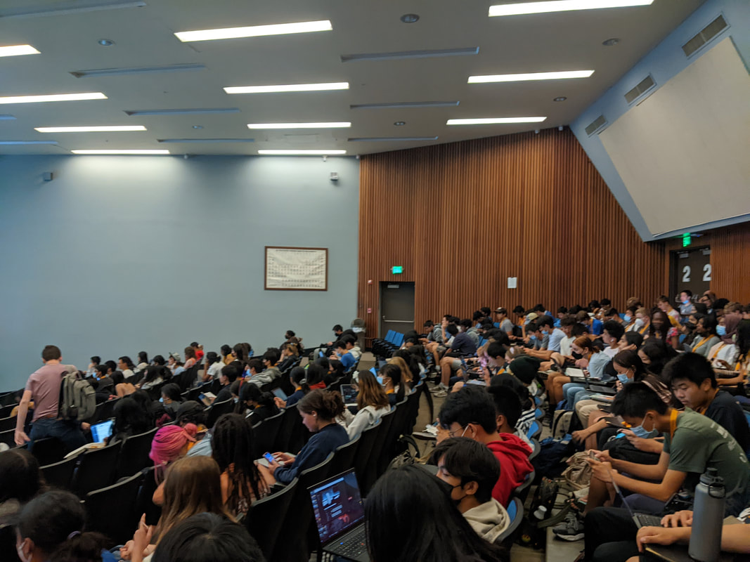
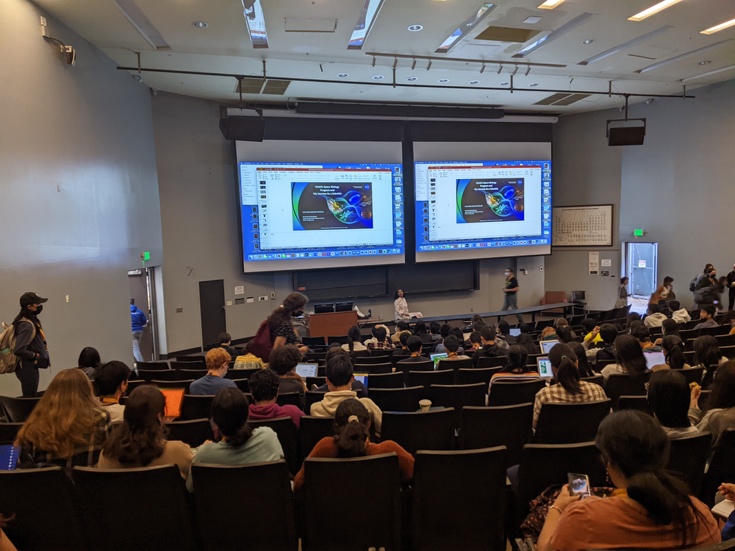
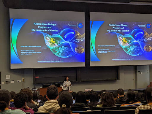
We kicked off the 4th week at COSMOS with NASA scientist, Prof. Sharmila Bhattacharya, and her fascinating, thought-provoking lecture on biology in space. She began with a thorough overview of deep space stressors, including microgravity, ionizing radiation, and altered temperature and atmosphere. These factors are necessary to consider when astronauts are spending extended periods of time drifting off in space and away from the comfort of Earth’s atmosphere.
She emphasized her research analyzing the biological responses of model organisms induced in space to construct an ideal, sustainable plant-human-microbial environment. We listened intently with admiration as she went on to describe the various experiments conducted at her NASA lab investigating the immunological and cardiovascular implications of space flight on such model organisms, including Drosophila (fruit flies). For example, in one particular study, her lab ran their trials within five experimental containers with fruit flies placed inside a rocket that followed a heliocentric orbit shortly after it launched. The data was transmitted back to a centralized computer and the results demonstrated space flight reduced phagocytosis efficiency in plasmatocytes (blood cells), meaning it hindered the ability of the host to fight bacterial and viral infections. Additionally, the effects were transgenerational, which means that they carried on past the original generation of fruit flies.
In another study, Prof. Sharmila Bhattacharya connected prolonged exposure to microgravity with cardiovascular defects and extracellular matrix (ECM) remodeling in-vivo. The gravity in space is significantly lacking compared to Earth, and the effects of such phenomena are believed to contribute to tissue remodeling and cardiac stress in Drosophila. Something particularly interesting that scientists still do not know the answer to is that human biological systems are typically impaired in space, while bacteria evolve to become more lethal. She noted the reason might be because viruses and bacteria are hard-wired to be pathogenic and their whole purpose is to become more virulent and capable of infiltrating the host.
In the end, she provided us with an inspiring story of her journey as a scientist and even a little about her experience with science policy as a Space Policy Advisor for the US Senate Committee in 2019. Currently, she is the program scientist for NASA’s Space Biology Program and has found a passion for teaching. I’m sure many of us admire her many accomplishments and extensive research experience in astrophysics and biology. Hopefully, we can all have a chance to make it to space in the near future.
- Hailey Van
Day 3: Blood Memory: Evaluating Antibody Responses to COVID-19 Infection
 Taken by Litong Deng.
On Wednesday morning, I walked into Classroom Unit 2 excitedly, took one look at the projector screen, and shouted an enthusiastic “yes!” It was the discovery lecture I had been looking forward to since COSMOS began. Prof. Rebecca DuBois was lecturing on COVID-19 antibodies.
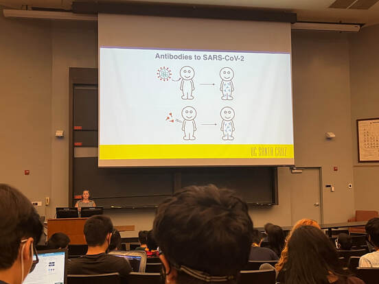
Prof. DuBois first began with a brief history of viruses other than SARS-CoV-2 (Severe Acute Respiratory Coronavirus 2, the virus that causes COVID-19) in the news. Then, she dove into a discussion about COVID-19 antibodies. She explained that they are Y-shaped protein molecules found throughout our blood — memories of the immune responses that we’ve had to infection and/or vaccination for diseases. When we are infected by a virus, white blood cells begin changing their genome to create different antibodies. When the correct antibody is produced, the white blood cell splits into a memory B cell, which ideally will duplicate itself for the rest of our lifetimes to give us lasting immunity, and a plasma cell, which produces more antibodies. These antibodies then bind to the surface of the viruses and prevent them from infecting our cells.
After antibodies, Prof. DuBois continued onto different COVID-19 vaccine technologies. We discussed mRNA vaccines (such as Pfizer-BioNTech, Moderna), Adenovirus-vectored vaccines (such as Johnson and Johnson, Oxford-AstraZeneca), and the “newest” technology, Novavax’s protein subunit vaccine. Although Novavax’s COVID-19 vaccine was only recently cleared for Emergency Use Authorization by the FDA, protein subunit vaccines have been used in many common vaccines, including flu and HPV vaccines. We learned that all authorized vaccines in the U.S. are trying to allow our cells to create spike proteins, so our body’s killer T cells can learn the appearance of SARS-CoV-2.
Now that we know that vaccines can produce antibodies, how can we detect antibodies? Prof. DuBois introduced us to serological testing, a way to detect antibodies. She introduced us to the two different types of antibody tests: nucleocapsid tests and spike tests. Nucleocapsid tests only look for evidence of past infection, whereas spike tests look for evidence of both past infection and past vaccination (but we won’t know which one).
Prof. DuBois concluded her talk with the technology that her lab has been developing — Biolayer Interferometry Immunosorbent Assay (BLI-ISA). BLI-ISA utilizes an instrument that measures how light comes down and back up through a biosensor. BLI-ISA can test for antibodies in less than 20 minutes and is more time-efficient than Enzyme-Linked Immunosorbent Assay (ELISA), another antibody testing method. Prof. DuBois explained that she has filed for Emergency Use Authorization from the FDA, but has not yet received a response.
Prof. DuBois answered many questions from the audience after her lecture and ended her presentation with a thunderous round of applause from the Cosmopolitan.
- Litong Deng